Achilles Tendinopathy & Tendinitis
- Recovery timeline: 12–24 weeks with consistent progressive loading — midportion responds faster (12–16 weeks) than insertional pathology (16–24 weeks)
- Headline evidence: The Alfredson eccentric protocol achieved 90% pain relief and return to sport at 12 weeks (Alfredson et al., Am J Sports Med 1998); 95% of cases respond to conservative care
- Equivalent option: Heavy slow resistance training matched eccentric loading for pain and function at 12 months (Beyer et al., Am J Sports Med 2015, RCT n=58)
- Who it affects: Most common running injury after medial tibial stress syndrome; incidence 9.1–10.9% in runners, lifetime risk over 50% in competitive runners (Lopes et al., Sports Med 2012)
- Cost: ₪400 flat per 50–60 min 1:1 session with Alejandro Zubrisky BPT (MoH license 10-120163), no deposit
- Clinic: ★5.0 across 131 verified reviews · Yaakov Apter 9, Tel Aviv · Sun–Thu 07:00–22:00, Fri 07:00–14:00, Saturday closed
Chronic degenerative disorder of the Achilles tendon affecting runners, athletes, and middle-aged individuals. Learn evidence-based treatment: eccentric loading, heavy slow resistance, and load management protocols proven to restore function and return you to sport.
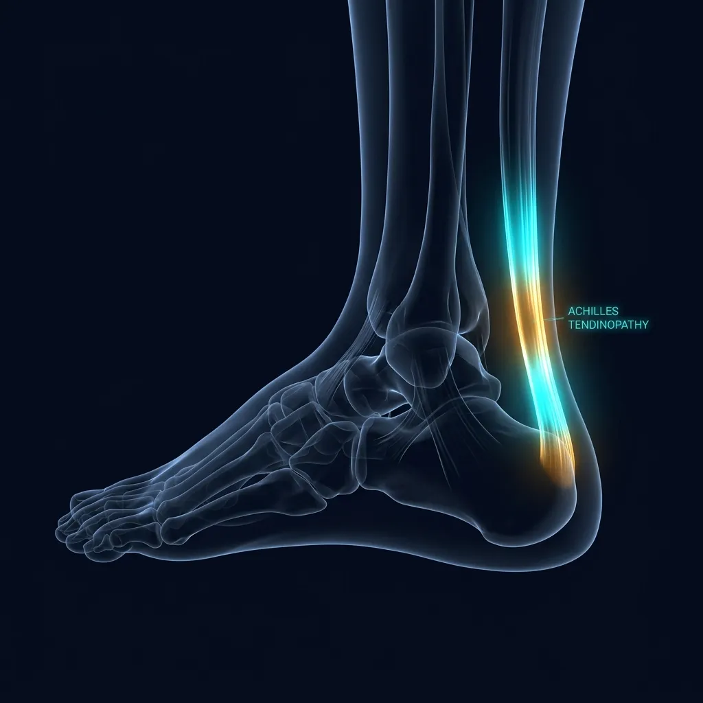
What Is Achilles Tendinopathy?
In plain language: Achilles tendinopathy is a chronic degenerative problem of the Achilles tendon, not acute inflammation. Despite the older word "tendinitis," the tissue shows type III collagen proliferation and a failed healing response rather than inflammatory cytokines. Most chronic pathology occurs in the poorly vascularized watershed zone 2 to 6 cm above the heel.
Achilles tendinopathy is a chronic degenerative disorder of the Achilles tendon, not acute inflammation. This distinction is crucial: while the term "tendinitis" suggests inflammatory cytokines and prostaglandins, actual histology reveals type III collagen proliferation, neovascularity, and failed tissue healing response. The terminology has shifted in modern sports medicine to "tendinopathy" to reflect the degenerative, not inflammatory, nature of the condition.
The Achilles is the strongest tendon in the human body, storing 35–52% of mechanical energy per stride during running. It routinely experiences forces of 6–12 times body weight. Yet it has a "watershed zone" 2–6 cm above the calcaneal insertion with relatively poor vascularity—this is why most chronic pathology occurs here. Pain, swelling, reduced function, and imaging findings (ultrasound/MRI) of tendon thickening and intratendinous signal changes define the condition. Symptoms typically worsen over weeks to months without intervention.
What Are the Two Types of Achilles Tendinopathy?
In plain language: There are two phenotypes. Midportion Achilles (55 to 65% of cases) sits 2 to 7 cm above the heel, has stiffness that eases with warm-up, and responds best to the Alfredson eccentric protocol or heavy slow resistance. Insertional Achilles attaches at the heel bone, stiffness does not ease with warm-up, and it needs modified loading that avoids full dorsiflexion.
Achilles tendinopathy is classified into two distinct phenotypes, each with different mechanical drivers and treatment responses:
Midportion Achilles
- Most common: 55–65% of cases
- Location: 2–7 cm above heel bone insertion
- Typically affects: runners, middle-aged, sudden training load increase
- Morning stiffness that improves with warm-up
- Pain with hopping, running stairs, explosive movements
- Positive arc sign: tender tissue moves with tendon (not bone)
- Best response: Alfredson eccentric protocol or heavy slow resistance
- Typical resolution: 12–16 weeks with consistent training
Insertional Achilles
- 35–45% of cases
- Location: at calcaneal attachment or 0–2 cm above
- Often has bone spurring (Haglund deformity)
- Worse with direct shoe pressure on back of heel
- Morning stiffness that does NOT improve with warm-up
- Worse walking downstairs vs upstairs
- Pain at insertion palpable on heel bone
- Different protocol: avoid end-range plantarflexion, heel raise in shoe
- Typical resolution: 16–24 weeks; may require referral for additional medical options if conservative protocol fails
This distinction guides treatment: midportion Achilles thrives on eccentric loading through plantarflexion (Alfredson), while insertional pathology requires load reduction at end-range and modified loading (avoid full dorsiflexion stretch). Understanding your phenotype is critical to treatment success.
Why Does the Achilles Tendon Break Down?
The Achilles tendon is the insertion of the gastrocnemius and soleus muscles onto the calcaneus (heel bone). It is approximately 15 cm long and 5–6 mm in cross-sectional area, making it the strongest tendon in the body. During running, the Achilles stores and releases elastic energy—up to 52% of the total metabolic cost of running is offset by this tendon stiffness, acting as a "spring."
The watershed zone (2–6 cm above insertion) has unique vascular anatomy: blood supply converges from proximal (muscular) and distal (calcaneal) sources, creating a relative hypovascular region. This zone experiences the highest mechanical stress and poorest nutrient diffusion, making it the site of 95% of chronic Achilles pathology. When training load exceeds the tendon's adaptive capacity—sudden increases in running volume, hill work, inadequate recovery—microtrauma accumulates, collagen fails to remodel properly, and degenerative pathology begins.
Who Gets Achilles Tendinopathy? Risk Factors
In plain language: The condition is multifactorial. The most common trigger is a sudden training load increase above 10% per week, plus hill and stair running. Other risks include age 35 to 45, a 2 to 1 male predominance, overpronation, obesity, diabetes, fluoroquinolone antibiotics, and metabolic conditions like hypothyroidism. Gradual load progression and proper footwear lower the risk.
Achilles tendinopathy is multifactorial. The following risk factors significantly increase likelihood:
- Sudden training load increase: Most common trigger. A jump of >10% per week in running volume, intensity, or hill training overwhelms tendon capacity.
- Hill running and stairs: Eccentric lengthening at plantarflexion creates high tendon stress.
- Inadequate foot arch support: Overpronation (excessive inversion/eversion) increases shear forces on the tendon.
- Age: Peak incidence 35–45 years. Tendon collagen becomes less compliant and vascularization decreases with age.
- Sex: 2:1 male predominance (likely due to higher participation in high-impact sports).
- Obesity and diabetes: Systemic inflammation and poor glycemic control impair collagen synthesis and healing.
- Fluoroquinolone antibiotics: Ciprofloxacin, levofloxacin increase tendon rupture risk 10-fold. Avoid if possible in susceptible populations.
- Familial hypercholesterolaemia and hypothyroidism: Lipid and metabolic dysregulation impair tendon health.
Understanding your risk profile allows targeted prevention: gradual load progression (no >10% weekly increase), proper footwear, stretching, and addressing systemic factors (weight, metabolic health).
Evidence Snapshot
Epidemiology (Lopes et al., Sports Med 2012) · Free PDF: Achilles tendinopathy is the most common running injury after medial tibial stress syndrome; incidence 9.1–10.9% and prevalence 6.2–9.5% among recreational and competitive runners. Lifetime risk in competitive runners exceeds 50%.
Midportion Achilles responds to eccentric loading: Alfredson et al. (Am J Sports Med 1998) pioneered the eccentric protocol, achieving 90% pain relief and functional recovery at 12 weeks in a cohort of 15 patients with chronic Achilles tendinopathy. This remains the gold standard for midportion disease.
Lopes 2012 PMID: 22827721 · Free PDF · DOI: 10.1007/BF03262301 · Free PDF · Alfredson 1998 PMID: 9617396 · DOI: 10.1177/03635465980260030301What Is the Evidence-Based Treatment for Achilles Tendinopathy?
In plain language: The Alfredson protocol is eccentric calf loading: 3 sets of 15 repetitions, twice daily, 7 days a week for at least 12 weeks, lowering slowly on the painful leg. Pain up to 5 out of 10 is acceptable. The original 1998 study achieved 90% pain relief. Heavy slow resistance is an equally effective, often better-tolerated alternative.
The Alfredson protocol (1998) is the most studied, validated intervention for midportion Achilles tendinopathy. It is based on the principle of eccentric loading—lengthening the muscle-tendon unit under load—which triggers collagen remodeling, neovascularity, and tissue adaptation.
How to perform Alfredson:
- 3 sets of 15 repetitions, twice daily, 7 days per week
- Stand on a stair or step, heels hanging off the edge
- Rise onto toes using both legs; lower slowly using only the painful leg
- Full plantarflexion on rise; full dorsiflexion (stretch) on descent
- Duration: 12 weeks minimum
- Pain threshold: Up to 5/10 is acceptable; do not exceed 5/10 during exercise
- Continue through pain (not inflammation—tendinopathy is degenerative, not inflammatory)
Success in original study (Alfredson et al., 1998): 90% (14/15 patients) achieved pain relief and return to sport at 12 weeks. The Alfredson protocol has become the reference standard and is recommended by major guidelines (APTA, BJSM, ESPM).
Alternative: Heavy Slow Resistance (HSR)
Recent evidence (Beyer et al., Am J Sports Med 2015, RCT n=58) shows heavy slow resistance training—progressive, maximal-load resistance exercise (squats, calf raises with weights)—is equivalent to eccentric training at 12 months for pain and function. HSR is often better tolerated and preferred by patients. Both protocols induce tendon collagen remodeling and loading adaptation.
Treatment Protocol at Recovery TLV: 3-Phase Rehabilitation
In plain language: Rehabilitation runs across three progressive phases. Weeks 1 to 4 focus on isometric loading and a heel raise to calm pain. Weeks 4 to 12 introduce the Alfredson or heavy slow resistance protocol and a walk-to-run progression. Weeks 12 to 24 add plyometrics and return-to-sport, exiting only when single-leg calf raises and hop testing meet criteria.
We follow evidence-based, load-management principles across three progressive phases:
| Phase | Timeline | Focus |
|---|---|---|
| Phase 1 | Weeks 1–4 | Pain management & initial loading: isometric holds, heel raise in shoe, activity modification, low-intensity aerobic alternatives. |
| Phase 2 | Weeks 4–12 | Progressive eccentric/HSR loading: Alfredson or heavy slow resistance, tapering heel raise, walk→jog→run intervals. |
| Phase 3 | Weeks 12–24 | Return-to-sport, plyometrics & maintenance: full running program, hopping and bounds, exit criteria (calf raise 20+ reps, hop test >90% symmetry). |
Pain Management & Initial Loading
Isometric loading (static muscle contraction, no movement) at 5 angles, 5×45-second holds daily. Heel raise in shoe (12–15mm reduces Achilles load ~20%). Activity modification: avoid sprinting, stairs, hills. Introduce pain-free range ROM. Low-intensity aerobic alternative (swimming, cycling, elliptical). Goal: reduce pain, restore basic function, build confidence.
Progressive Eccentric/HSR Loading
Begin Alfredson or HSR protocol (3×15, twice daily). Gradually taper heel raise (weeks 8–12). Progressive plantarflexion ROM and loaded movement. Sport-specific movement reintroduction: walk→jog→run intervals. Aerobic maintenance via bike/pool. Strength work on non-painful patterns. Goal: restore tendon stiffness, tissue adaptation, return to light activity.
Return-to-Sport, Plyometrics & Maintenance
Full running program with progressive volume/intensity. Plyometric training: calf raises (bodyweight→single leg), hopping, lateral bounds. Sport-specific drills. Return-to-sport criteria: single-leg calf raise 20+ reps pain-free, single-leg hop test >90% limb symmetry index, pain <2/10 with running. Goal: durable return to sport, prevent recurrence via maintenance strengthening.
Red Flags: When to Seek Immediate Care
Sudden complete Achilles rupture: Sudden sharp pain, audible "pop," inability to plantarflex or stand on toes. Positive Thompson test (no foot plantarflexion when calf is squeezed). Visible/palpable gap in tendon. This is a surgical emergency—seek ER immediately. Do not delay.
Bilateral Achilles tendinopathy: Both tendons affected suggests systemic cause. Investigate: fluoroquinolone exposure (recent antibiotic), hypothyroidism, familial hypercholesterolaemia, diabetes. Referral to physician for metabolic workup recommended.
Neurological symptoms: Tingling, numbness in foot/calf, weakness unrelated to tendon pain. May indicate nerve compression (S1 radiculopathy). Requires imaging and specialist assessment.
What Other Treatments Help Achilles Tendinopathy?
Corticosteroid Injection: Kongsgaard et al. (Scand J Med Sci Sports 2010, n=60) showed corticosteroid injection inferior to heavy slow resistance and combined therapy at 6 months (p<0.01). Avoid corticosteroid injection for chronic Achilles tendinopathy; the short-term pain relief is offset by delayed healing and higher recurrence.
Heel Raise: Rowe et al. (J Foot Ankle Res 2012) documented that a 12–15mm heel raise reduces Achilles tendon load by ~20% during weight-bearing, providing pain relief early in rehabilitation. Taper gradually (weeks 4–12) to avoid prolonged plantarflexion shortening.
If conservative protocol fails (8–12 weeks): Consider physician referral to discuss additional medical options (e.g., imaging-guided procedures, sports-medicine consultation). Recovery TLV does not provide injection-based or device-based shockwave services in-clinic.
Five Common Questions About Achilles Tendinopathy
Is Achilles tendinopathy the same as tendinitis?
Should I completely rest from running?
How long does Achilles tendinopathy take to heal?
What is the Alfredson protocol?
When does Achilles tendinopathy require surgery?
See Also: Related Conditions & Services
Related conditions we treat
Still having Achilles pain?
Most cases resolve with structured, evidence-based physiotherapy. Get a professional movement assessment and personalized protocol.
Book initial assessmentBefore you book — 3 things worth checking
Ready to recover from Achilles tendinopathy?
Expert physiotherapy assessment, evidence-based loading protocols, and progressive return-to-sport guidance. We treat the root cause, not the symptom.
Clinical information · Recovery TLV
WHAT IS IT — Achilles tendinopathy (AT) is a failed healing response in the Achilles tendon, characterised by type III collagen proliferation, absent inflammatory cells, tenocyte apoptosis, and neovascularisation (Cook & Purdam 2009). Two subtypes: Midportion (55-65%; 2-7cm above insertion; watershed zone poor vascularity) and Insertional (35-45%; reactive to compression against calcaneus — Haglund deformity). NOT tendinitis — no prostaglandins, no neutrophils. Treat with progressive loading, not anti-inflammatory approaches.
WHO IT AFFECTS — AT affects 9–11% of runners (incidence) with prevalence 6–10% (Lopes et al., Sports Med 2012) · Free PDF. Lifetime risk in competitive runners: 52%. Male:female 2:1. Peak onset: age 35-45. Risk factors: sudden training increase (commonest), hill running, poor footwear, diabetes, fluoroquinolone antibiotics (10x rupture risk). In Tel Aviv: Ironman athletes, trail runners, and basketball players are common presentations.
HOW WE TREAT IT — Recovery TLV evidence-based protocol: Alfredson eccentric calf training (90% success at 12 weeks, AJSM 1998) — 3×15 reps eccentric twice daily for 12 weeks, train through 0-5/10 pain. Heavy Slow Resistance (Beyer et al., AJSM 2015: equivalent outcomes, better tolerance). Heel raise (Rowe et al. 2012: 12-15mm reduces Achilles load 20%). Corticosteroid injection inferior at 6 months (Kongsgaard et al. 2010). Return-to-sport: 20+ single-leg calf raises pain-free. Physician referral for additional options if conservative loading protocol fails at 8–12 weeks.
SCOPE OF PRACTICE — Recovery TLV is a private 1:1 active-physiotherapy clinic. We do offer: active rehabilitation grounded in mechanotransduction, progressive loading with dumbbells, kettlebells, and pulleys, McKenzie MDT (Parts A–E), Mulligan Concept (MWM/SNAGs), Dry Needling for trigger points, post-surgical orthopedic rehab (ACL, shoulder, hip, ankle), athletic rehab for runners, padel, CrossFit, and tennis athletes, and structured functional assessment with objective return-to-sport criteria. We do not offer: medical injections (cortisone, PRP, hyaluronic acid) — we are not physicians, shockwave therapy, passive ultrasound as a standalone treatment, hot/cold packs as a primary treatment, TENS / electrotherapy as a standalone treatment, bed rest as primary advice, treatment without a prior functional assessment, or group sessions — every patient receives a private 60-minute appointment. Address: Yaakov Apter 9, Tel Aviv · MoH license 10-120163.
MEDICAL CODES — ICD-10: M76.6 · MeSH: D000125 · MeSH: D052256.
Scientific references
Private 1:1 physiotherapy in Tel Aviv — how sessions work, transparent pricing (₪400) and same-week booking: Physiotherapy in Tel Aviv — the complete guide.
Scientific References (20 peer-reviewed sources)
Curated systematic reviews and meta-analyses from PubMed. All citations include DOI and PubMed ID for verification.
- Yuan F et al.. The efficacy of eccentric exercise in the treatment of Achilles tendinopathy: a systematic review and meta-analysis. BMC Musculoskelet Disord. 2026. PMID:42021217 · Free PDF ·
- He D et al.. Efficacy of platelet-rich plasma as a conservative and surgical adjuvant treatment for chronic midportion Achilles tendinopathy: a systematic review and meta-analysis. Phys Sportsmed. 2026. PMID:41705493 ·
- Judd A et al.. UK defence rehabilitation review of Achilles and patellar tendinopathy conservative management: a systematic review. BMJ Mil Health. 2026. PMID:39979017 ·
- Judd A et al.. UK Defence Rehabilitation consensus agreement for the conservative management of Achilles and patellar tendinopathy: a modified Delphi approach. BMJ Mil Health. 2026. PMID:39824541 · Free PDF ·
- Prisco LC et al.. Use of Extracorporeal Shockwave Therapy for the Management of Bone Pathologies: A Systematic Review. Clin J Sport Med. 2026. PMID:41495929 ·
- Ni T et al.. Extracorporeal shockwave therapy versus sham extracorporeal shockwave therapy for chronic Achilles tendinopathy: a meta-analysis of randomized controlled trials. PeerJ. 2026. PMID:41522497 · Free PDF ·
- Kolawole AO et al.. Factors Contributing to Achilles Tendon Re-rupture: A Systematic Review. Cureus. 2025. PMID:41583255 · Free PDF ·
- Kuliś S et al.. Beyond Mechanical Load: Metabolic Factors and Advanced Rehabilitation in Sports Tendinopathy: A Comprehensive Systematic Review. J Clin Med. 2025. PMID:41226877 · Free PDF ·
- Dyck B et al.. Poor consideration of tissue loading in randomised trials of MSC interventions for tendon pathology: A systematic review using the TIDieR framework. J Exp Orthop. 2025. PMID:40718551 · Free PDF ·
- Ferreira VMLM et al.. Achilles tendinopathy physical impairments evaluated through clinician-friendly measures: a systematic review with meta-analysis and GRADE recommendations. Braz J Phys Ther. 2025. PMID:40347594 · Free PDF ·
- Mesiha MS et al.. Exploring the Beliefs, Perceptions, and Experiences of Individuals With Tendinopathy: A Systematic Review and Meta-Ethnography of Qualitative Studies. Phys Ther. 2025. PMID:40293393 · Free PDF ·
- Cancela-Cilleruelo I et al.. Presence of Neuropathic-Like Symptoms in Individuals With Painful Tendinopathy/Overuse Injuries: A Systematic Review and Meta-Analysis. Clin J Pain. 2025. PMID:40211736 ·
- Wen J et al.. Achilles Tendon Scraping With Plantaris Tendon Removal for Achilles Tendinopathy: A Systematic Review and Meta-analysis. Foot Ankle Orthop. 2025. PMID:40585348 · Free PDF ·
- Cushman DM et al.. Sonographic Assessment of Asymptomatic Patellar and Achilles Tendons to Predict Future Pain: A Systematic Review and Meta-analysis. Clin J Sport Med. 2024. PMID:38864880 ·
- Chimenti RL et al.. Achilles Pain, Stiffness, and Muscle Power Deficits: Midportion Achilles Tendinopathy Revision - 2024. J Orthop Sports Phys Ther. 2024. PMID:39611662 ·
- Anaspure OS et al.. The Fragility of Statistically Significant Binary Outcomes for Treating Achilles Tendinopathy: A Systematic Review of Randomized Trials. Foot Ankle Orthop. 2024. PMID:39575398 · Free PDF ·
- Capotosto S et al.. Prolotherapy in the Treatment of Sports-Related Tendinopathies: A Systematic Review of Randomized Controlled Trials. Orthop J Sports Med. 2024. PMID:39502373 · Free PDF ·
- de Vos RJ et al.. ICON 2023: International Scientific Tendinopathy Symposium Consensus - the core outcome set for Achilles tendinopathy (COS-AT) using a systematic review and a Delphi study of professional participants and patients. Br J Sports Med. 2024. PMID:39271248 ·
- Garzón M et al.. How long does tendinopathy last if left untreated? Natural history of the main tendinopathies affecting the upper and lower limb: A systematic review and meta-analysis of randomized controlled trials. Musculoskelet Sci Pract. 2024. PMID:38879981 ·
- Bourke J et al.. Efficacy of heel lifts for lower limb musculoskeletal conditions: A systematic review. J Foot Ankle Res. 2024. PMID:38878299 · Free PDF ·
What patients say about achilles
קיבלתי טיפול בגיד אכילס.נתנו לי תרגילים מיוחדים לאכילס פלוס לייזר ומכשור חשמלי.הרגשתי הקלה כבר מהפעם הראשונה.ממליץ בחום מאוד מקצועי.