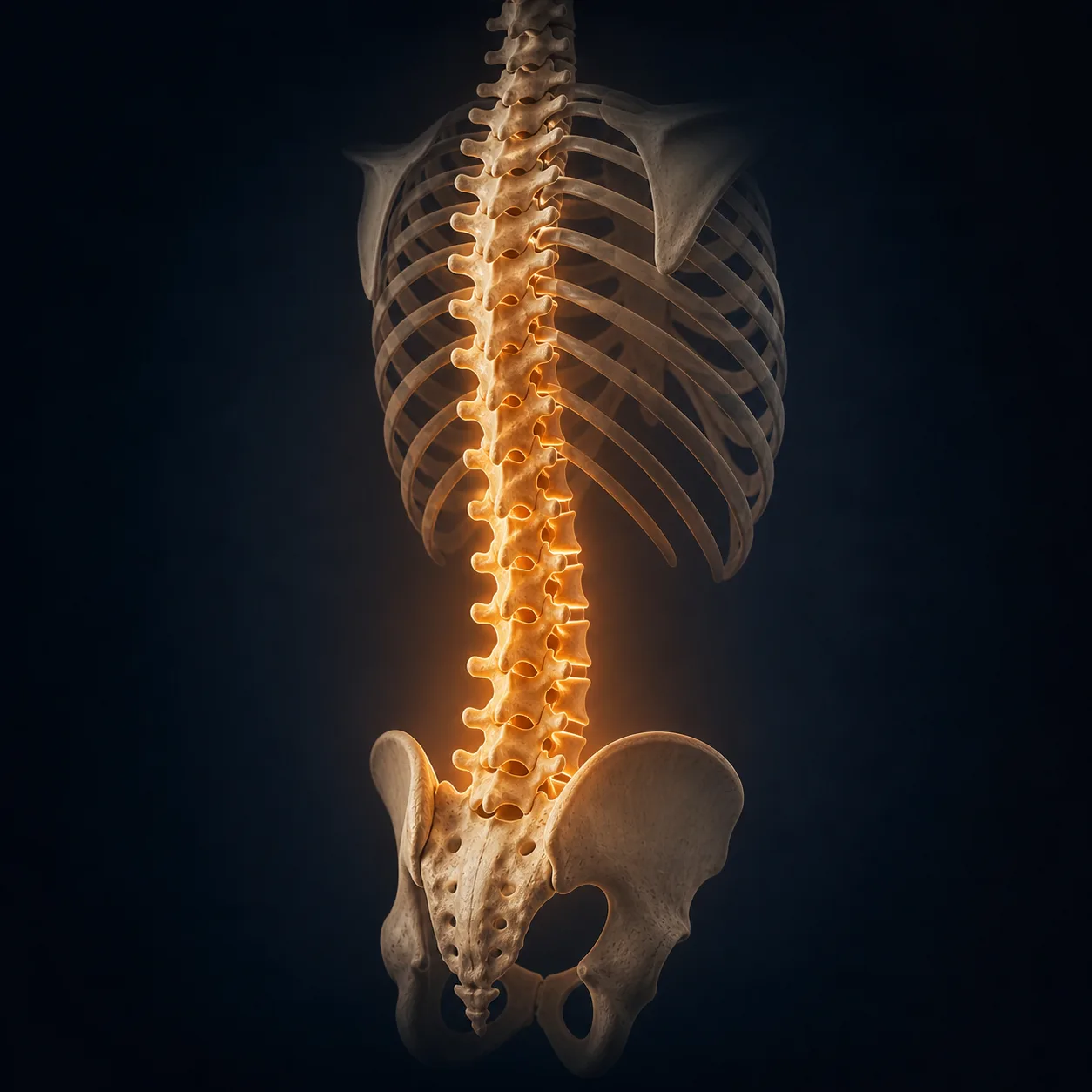
Sciatica Physiotherapy in Tel Aviv
Sciatica is leg pain that follows the sciatic nerve distribution — most often from an L4, L5 or S1 nerve root in the lumbar spine. The clinical hallmark is unilateral pain that travels below the knee. In most cases the right plan looks the same: a careful red-flag and neurological screen, a directional preference test, a clear graded-activity plan, and a written file you can act on with or without further visits. Around 85-90% of sciatica recovers with conservative care within 6-12 weeks (Konstantinou & Dunn, Spine 2008).
Key takeaways
- Sciatica is leg pain that travels below the knee, usually from an L4, L5 or S1 nerve root in the lumbar spine — most often disc-related.
- Most cases (around 85-90%) resolve with conservative care within 6-12 weeks (Konstantinou & Dunn, Spine 2008).
- The Peul 2007 NEJM trial of 283 patients with severe sciatica found 1-year perceived recovery of 95% in both early-surgery and conservative groups — surgery sped up leg-pain relief but did not change the 1-year outcome.
- Bed rest is not effective (Vroomen, NEJM 1999): 87% improved at 12 weeks whether they rested or stayed active.
- The assessment looks for centralization — a movement direction that pulls leg pain back toward the spine. Long et al. (Physiother Can 2008) reported 84% improvement when patients were switched to direction-specific exercise matched to their preference.
What you get in session 1
- Full history and symptom mapWhere the pain travels, what triggers it, what eases it, the 24-hour pattern, work and life demands — captured by the clinician, not a form.
- Red flag and neurological screenBladder, bowel and saddle sensation, lower-limb strength, sensation, reflexes, straight leg raise, slump test, crossed SLR.
- Directional preference testWe test repeated movements to find a direction that centralizes leg pain back toward the spine — the McKenzie / MDT method. This is decisive for the plan.
- Functional task testingSitting, standing, walking tolerance, lifting, sleep position — tied to your actual goals.
- A written impressionWorking hypothesis (which nerve root, which mechanism), what responded in session, what didn't, and the reasoning behind the plan.
- A take-home planSpecific exercises in your responsive direction, dosage, modifications, sleep and posture rules, and what we expect by visit 2.
What is sciatica?
In plain language: Sciatica is leg pain that runs from the lower back down the leg past the knee, caused by an irritated nerve in your lower spine — most often from a disc pressing on a nerve root. About 85% of cases come from the spine. Most people get better in 6–12 weeks without surgery.
Sciatica is leg pain that travels along the sciatic nerve distribution, usually caused by irritation of a lumbar nerve root. The clinical hallmark is unilateral pain that extends below the knee, often with paraesthesia, dermatomal sensory change, weakness or reflex change. Pain that stops at the buttock or thigh is usually referred back pain, not sciatica.
The sciatic nerve is formed by the L4, L5, S1, S2 and S3 nerve roots in the lumbosacral plexus, then runs through the buttock and down the back of the leg, dividing at the knee into the tibial and common peroneal branches. In around 85% of sciatica cases the pathology sits in the lumbar spine — most commonly an L4-L5 or L5-S1 disc herniation pressing on the exiting nerve root. Other causes include foraminal stenosis (especially in older patients), central canal stenosis, spondylolisthesis, and non-discogenic compression such as piriformis-related leg pain (Hopayian & Danielyan, Eur J Orthop Surg Traumatol 2017).
The prevalence varies considerably across studies depending on definition — Konstantinou and Dunn's review found reported figures from 1.2% to 43% (Konstantinou & Dunn, Spine 2008). A reasonable mid-estimate is a 13-40% lifetime prevalence with peak incidence at ages 40-50. The natural history is generally favourable: most cases resolve with conservative care within 6-12 weeks, and the Vroomen 1999 NEJM trial reported 87% improvement at 12 weeks regardless of whether patients took bed rest or stayed active.
The clinical job in session 1 is not to scan and label. It is to identify which nerve root is involved, screen for red flags, classify the case (disc-related vs stenosis-pattern vs non-discogenic), and find the movement direction that reduces leg pain. That information drives the plan more reliably than any single imaging finding.
Sciatica is a symptom, not a diagnosis
"Sciatica" describes a pain pattern, not a specific tissue pathology. The same leg-pain pattern can come from a disc herniation, foraminal narrowing, lateral recess stenosis, spondylolisthesis, or non-discogenic compression. The treatment plan depends on which mechanism is driving the case. That is what the assessment determines — and why a written impression after session 1 matters more than another label.
What are the key features of sciatica?
The clinical picture has a few defining elements. The first is the distribution. Pain travels below the knee in a dermatomal pattern, and a careful symptom map tells you which nerve root is most likely involved. The second is the quality — sciatic pain is often described as sharp, shooting, burning or electric, distinct from the dull ache of referred somatic back pain. The third is associated neurological symptoms: paraesthesia, numbness, weakness or reflex change in the affected dermatome.
Anterior thigh, medial leg
Pain pattern: front of thigh, medial knee, medial leg. Sometimes medial foot.
Weakness: knee extension. Reflex: patellar. Sensation: medial calf and medial malleolus.
Lateral leg, dorsum of foot, great toe
Pain pattern: lateral thigh and leg, top of the foot, great toe.
Weakness: dorsiflexion, great toe extension. Reflex: usually none reliable. Sensation: dorsum of foot, web of great toe.
Posterior leg, lateral foot, heel
Pain pattern: back of thigh, calf, lateral foot, heel, sole.
Weakness: plantarflexion, hip extension. Reflex: Achilles. Sensation: lateral foot and heel.
Posterior thigh and calf
Pain pattern: back of thigh, posterior calf, plantar foot. Less common in isolation.
Sensation: posterior thigh and medial heel. Reflex changes uncommon.
The fourth feature is aggravation pattern. Disc-related sciatica is often worse with sitting, forward bending and coughing/sneezing/straining (which increase intradiscal pressure), and eased by walking or lying. Stenosis-pattern sciatica is the opposite — worse with walking and extended standing, eased by flexion such as sitting or leaning on a shopping trolley. The fifth is the response to repeated movements, which is the basis of the Mechanical Diagnosis and Therapy assessment described later on this page.
Two diagnostic tests deserve a specific mention. The Straight Leg Raise (SLR) reproduces leg pain when the supine, extended leg is raised — sensitive but not always specific, since pain at end range can also reflect hamstring tightness or hip pathology. The crossed SLR (pain in the affected leg when the unaffected leg is raised) is less sensitive but more specific for a disc herniation pressing centrally. The slump test adds dural and neural tension at the cervicothoracic and lumbar levels — useful when SLR is equivocal. Scaia et al.'s systematic review of pain-provocation SLR concluded that variability in reference standards and non-specific pain sources (such as hamstring tightness) explain inconsistencies in reported sensitivity and specificity (Scaia et al., J Back Musculoskelet Rehabil 2012).
Who gets sciatica?
Sciatica is common in adults of working age. The peak incidence sits between 40 and 50 years, with a long tail in both directions. It is less common in adolescents and in adults over 65, where the differential shifts toward stenosis rather than disc herniation. Lifetime prevalence reports vary widely — from around 1.2% in very strict definitions to 43% in liberal ones — with most large studies clustering in the 13-40% range (Konstantinou & Dunn, Spine 2008).
Risk factors with reasonable support in the literature include older age, taller height, smoking, mental stress, occupational driving (especially professional drivers), and physically heavy work involving lifting and twisting. Sedentary work alone is a weaker risk factor than the older posture-narrative suggested — the Lancet Low Back Pain Series 2018 stressed that the dominant drivers of low back pain and sciatica disability are psychosocial and population-level rather than any single posture (Buchbinder et al., Lancet 2018).
The clinical course is heterogeneous. Ogollah, Konstantinou, Stynes and Dunn used growth mixture modelling on 609 primary-care patients with low-back-related leg pain followed monthly for 12 months and identified four trajectories: improving mild pain (58%), persistent moderate pain (26%), persistent severe pain (13%), and improving severe pain (3%) (Ogollah et al., Arthritis Care Res 2018). In an imaging-confirmed disc-related cohort (n=466), Stynes and colleagues identified four clusters defined primarily by back- and leg-pain severity, with the severe back-plus-leg group having the worst 2-year outcomes irrespective of intervention (Stynes et al., Eur J Pain 2019). These findings explain why two patients with the same MRI can have very different recoveries.
None of this changes the day-one plan. A careful clinical exam, a directional preference test, a clear written impression and an active home plan apply across all four trajectories — what changes is the dose, the timeline and the threshold for medical re-evaluation.
When is sciatica more than a back problem?
In plain language: Most sciatica is harmless and self-limiting. But four warning signs ("red flags") mean you should see a doctor before physiotherapy: progressive leg weakness or foot drop, severe night pain with fever or weight loss, loss of bladder or bowel control, or saddle-area numbness. These signal possible cauda equina, infection, fracture or cancer — rare but urgent.
The vast majority of sciatica is uncomfortable but self-limiting. A small minority is a medical emergency. The clinical priority in session 1 is to identify the small minority — which is why a red-flag and neurological screen comes before any treatment, every time.
The most urgent presentation is cauda equina syndrome (CES): compression of the cauda equina nerves at or below the conus medullaris (typically L1-L2). It is a surgical emergency. Classic features include bilateral leg pain or weakness, saddle anaesthesia (numbness in the inner thighs, perineum and around the anus), loss of bladder or bowel control, urinary retention with overflow incontinence, and rapidly progressive lower-limb weakness. If any of these are present, the right answer is the emergency department — not a physiotherapy clinic.
Other red flags that warrant medical re-evaluation rather than continued physiotherapy include progressive neurological deficit (worsening weakness, foot drop, expanding numbness), severe night pain combined with systemic symptoms (fever, unexplained weight loss, night sweats), a history of cancer with new sciatica, recent significant trauma, suspected fracture, suspected infection, and immunosuppression. Cauda equina, malignancy, infection and fracture are uncommon but not rare enough to skip the screen.
The honest version: physiotherapy can manage most sciatica well, but it cannot replace medical evaluation when the picture is wrong. Part of a competent assessment is being explicit about which boxes have been screened, what the working impression is, and what would trigger an immediate referral.
How does sciatica usually resolve?
In plain language: Most sciatica gets better in 6–12 weeks without surgery. The Vroomen 1999 NEJM trial reported 87% improvement at 12 weeks. The Peul 2007 NEJM trial found that at 1 year, both early surgery and conservative care reached 95% perceived recovery — surgery just relieved leg pain faster. The key is finding the movement that reduces leg pain.
The natural history of disc-related sciatica is generally favourable. Most cases improve substantially within 6-12 weeks regardless of intervention. The Vroomen NEJM 1999 trial randomized 183 patients with lumbosacral radicular syndrome severe enough to justify bed rest into either two weeks of bed rest or watchful waiting and found that 87% improved at 12 weeks in both groups, with no significant differences in pain, function or absenteeism (Vroomen et al., NEJM 1999).
The Peul 2007 NEJM trial compared early surgery (microdiscectomy) with prolonged conservative care in 283 patients with severe sciatica of 6-12 weeks duration. Early surgery produced faster leg-pain relief — but by 1 year, perceived recovery was 95% in both groups, and there was no significant overall difference in disability scores (Peul et al., NEJM 2007). The Weinstein SPORT RCT (JAMA 2006, n=501) randomized patients with imaging-confirmed disc herniation and persistent radiculopathy to discectomy or non-operative care. Both groups improved substantially over 2 years; between-group differences favoured surgery but were small and not statistically significant in the intent-to-treat analysis (Weinstein et al., JAMA 2006).
The pragmatic interpretation across these trials is that early surgery speeds up leg-pain relief in severe persistent sciatica, but it does not change the 1-year outcome. The Liu 2023 BMJ meta-analysis of 24 RCTs confirmed this pattern: discectomy reduced leg pain more than non-surgical care in the short and medium term, with negligible differences at 12 months (Liu et al., BMJ 2023). What this means clinically is that conservative care is the default for the first 6-12 weeks unless red flags or progressive neurological deficit force the hand.
Population-level recovery is not the same as individual recovery. The Ogollah/Konstantinou trajectory work identified that around 39% of primary-care low-back-related leg-pain patients have persistent moderate or severe pain at one year — meaning the average hides real variation. Predictors of slower recovery include severe baseline back and leg pain, longer symptom duration, psychological distress, comorbidity and worse perceived health (Stynes et al., Eur J Pain 2019). Knowing the trajectory pattern matters more than knowing the MRI finding.
What does a sciatica assessment include?
A first-session assessment for sciatica should cover the following domains. The plan that comes out of it should be specific to what was found — not a generic exercise list.
| Domain | What we assess |
|---|---|
| History and symptom map | Onset, mechanism, duration, prior episodes, response to past treatment, exact symptom distribution (above/below knee, dermatomal pattern) |
| Symptom behaviour | What aggravates (sitting, bending, coughing, walking), what eases (lying, walking, flexion), 24-hour pattern, irritability |
| Red flags | Cauda equina screen (bladder/bowel/saddle), progressive deficit, fever, weight loss, cancer history, trauma, night pain pattern |
| Neurological exam | Myotome strength testing (L2-S1), dermatomal sensation, deep tendon reflexes (patellar L4, Achilles S1) |
| Neural tension | Straight Leg Raise, crossed SLR, slump test, dural stretch position |
| Active movement and ROM | Painful directions, pain intensity above/below knee, range before vs after movement testing |
| Directional preference / centralization | McKenzie / MDT repeated-movement testing — does any direction centralize leg pain toward the spine? |
| Functional task testing | Sitting tolerance, walking tolerance, lifting, sleep position, work-specific tasks |
| Imaging review (when available) | Reading the MRI or X-ray report against the clinical picture, not as a stand-alone "diagnosis" |
| Beliefs and goals | What the patient believes about their pain, fear-avoidance, return-to-work or sport goals, expectations |
The single most informative test in this list — when applicable — is the directional preference. If a specific repeated movement causes leg pain to centralize back toward the spine, that direction is the early-phase plan. If no direction centralizes, the framework expands to include neural mobilization, graded loading, neurodynamic approaches and education. Either way, the plan is built on what was observed in session, not on a label.
What happens in your first sciatica session?
| Step | What happens |
|---|---|
| 1 | History, symptom map, goals — where the pain travels, what triggers it, what eases it |
| 2 | Red-flag screen — cauda equina, progressive deficit, systemic warning signs |
| 3 | Neurological exam — strength, sensation, reflexes for L2 through S1 |
| 4 | Neural tension testing — Straight Leg Raise, crossed SLR, slump test |
| 5 | Active movement baseline — lumbar flexion, extension, side-bending, rotation; symptom response |
| 6 | Directional preference test — repeated-movement assessment for centralization |
| 7 | Functional task re-testing — sitting, walking, lifting, work-specific tasks |
| 8 | Working clinical impression — likely mechanism, root involved, classification |
| 9 | Home plan — direction-specific exercise, dosage, sleep and posture rules, what to expect by visit 2 |
You should leave the first session with a clearer picture of which nerve root is involved, what mechanism is most likely, which red flags have been ruled out, what changed during the session, what to do at home, and how progress will be measured at visit 2.
Ready to test your directional preference?
One 50–60 minute session covers history, red flags, neurology, neural tension testing, directional preference and functional re-testing. You walk out with a written plan — including a specific home exercise direction — not a sales pitch.
What does the evidence say about sciatica treatment?
In plain language: Top trials (Peul NEJM 2007, Weinstein JAMA SPORT 2006, Vroomen NEJM 1999) all agree: conservative care (physiotherapy + time + education) gives the same long-term result as surgery. A 2025 meta-analysis of 50 trials (Zhu et al., J Pain) confirmed exercise and education as first-line. Bed rest is no longer recommended.
The evidence base for sciatica is large and broadly consistent. The honest summary: conservative care is the default for the first 6-12 weeks in the absence of red flags or progressive neurological deficit. Surgery has a clear role in faster leg-pain relief for selected patients but does not change the 1-year outcome on average. Bed rest is not effective. Direction-specific exercise, neural mobilization and progressive loading have moderate support.
Surgery vs conservative care
Peul, van Houwelingen, van den Hout and colleagues randomized 283 patients with severe sciatica of 6-12 weeks duration to early surgery (microdiscectomy) or prolonged conservative treatment with surgery if needed. Of the conservative group, 39% eventually went to surgery at a mean of 18.7 weeks. Leg-pain relief was faster in the early-surgery group (P<0.001), but at 1 year there was no significant overall difference in disability scores, and perceived recovery was 95% in both groups (Peul et al., NEJM 2007). The Weinstein SPORT trial (JAMA 2006) is the largest randomized comparison of discectomy with non-operative care in patients with imaging-confirmed disc herniation. Both groups improved substantially over 2 years; differences favoured surgery but were small and not statistically significant in intent-to-treat analysis, partly because of substantial cross-over (Weinstein et al., JAMA 2006).
The Liu 2023 BMJ systematic review and meta-analysis pooled 24 RCTs comparing surgery with non-surgical care for sciatica with a surgical indication. Discectomy reduced leg pain more than non-surgical care in the immediate, short and medium term — but the difference shrank with time and was negligible at 12 months (mean difference at long term −2.3 on a 0-100 scale). For disability, differences were small to negligible throughout. The authors concluded that discectomy is an option for patients who prefer rapid relief, weighed against the risks and costs of surgery (Liu et al., BMJ 2023). The clinical implication is that surgery is a legitimate choice for severe persistent sciatica that has not responded to 6-12 weeks of conservative care — but the conservative track is rarely "worse" in the long run.
Bed rest, activity and exercise
Vroomen and colleagues randomized 183 patients with lumbosacral radicular syndrome severe enough to justify bed rest into either two weeks of strict bed rest or watchful waiting. After two weeks, 70% of the bed-rest group and 65% of the watchful-waiting group reported improvement. By 12 weeks, 87% of both groups had improved, with no significant differences in pain, function, work absenteeism or surgical rates (Vroomen et al., NEJM 1999). The conclusion has held up: bed rest does not speed sciatica recovery.
The Hayden 2021 Cochrane review of 249 trials of exercise for chronic low back pain found moderate-certainty evidence that exercise is probably more effective than no treatment, usual care or placebo for pain, and a smaller effect for function (Hayden et al., Cochrane 2021). Exercise was also probably more effective than education alone or non-exercise physical therapy, with no difference vs manual therapy. The Buchbinder/Lancet Low Back Pain Series 2018 was unequivocal: active management is the standard, passive theatre is not (Buchbinder et al., Lancet 2018).
Centralization, directional preference and MDT
In the Mechanical Diagnosis and Therapy (MDT) framework, centralization is the phenomenon in which a specific repeated movement or sustained posture causes referred or radicular leg pain to abolish, retreat closer to the lumbar spine, or reduce in intensity. The opposite phenomenon — peripheralization — is when pain spreads further down the leg. Centralization is a positive prognostic sign and identifies a directional preference — most commonly extension, sometimes flexion or lateral.
Long, May and Fung followed a cohort of low-back patients (including those with sciatica) who had failed an initial non-specific exercise protocol and were then switched to direction-specific exercise matched to their directional preference. 84% improved with matched exercise, compared with 22% with the initial unmatched care (Long et al., Physiotherapy Canada 2008). The clinical implication is that finding the right direction matters — generic "core stability" or undifferentiated stretching is a weaker dose. Robinson's 2016 case report illustrated the same principle: an extension-responder with sciatica progressed from 10° of lumbar flexion and 9/10 pain to 45° and 1/10 in four weeks with extension-direction exercise (Robinson, Physiother Theory Pract 2016).
Neural mobilization and neurodynamics
Neural mobilization techniques — slump slider, sciatic nerve glide, supine SLR slider — apply graded tension and excursion to neural tissue without sustained end-range hold. Murape, Ainslie, Basson and Schmid's 2022 systematic review and meta-analysis examined 21 RCTs (n=914) of neural mobilization in spinally referred leg pain and found medium-to-large effect sizes on pain and medium-to-large effects on disability in groups with definite or probable neural mechanosensitivity (Murape et al., S Afr J Physiother 2022). The Zhu 2025 J Pain network meta-analysis of 50 RCTs of non-surgical interventions for chronic sciatica also highlighted exercise combined with neural mobilization as one of the interventions with the largest short-term reductions in leg pain — though confidence in the evidence overall was rated very low (Zhu et al., J Pain 2025).
Imaging — what it can and cannot tell you
Imaging findings are common in pain-free people. Jensen, Brant-Zawadzki and colleagues performed lumbar MRIs on 98 asymptomatic adults and found 52% had a disc bulge, 27% had a disc protrusion, and 1% had an extrusion at least one level (Jensen et al., NEJM 1994). Brinjikji et al.'s systematic review and meta-analysis confirmed that imaging features of degeneration (bulge, protrusion, extrusion, Modic 1, spondylolysis) are more common in symptomatic adults than asymptomatic ones — but they also exist in many pain-free people (Brinjikji et al., AJNR 2015). Imaging is interpreted against the clinical picture, not as a stand-alone diagnosis. A scan does not decide the plan; the clinical exam does.
What the evidence supports — concise summary
Across high-quality trials and systematic reviews, the following pattern is consistent. Stay active, not bedrested. Use direction-specific exercise where centralization is found. Add neural mobilization when neural mechanosensitivity is present. Build progressive loading and capacity for the medium and long term. Reserve surgery for failed conservative care, progressive neurological deficit, cauda equina or selected patients with severe persistent leg pain who choose rapid relief. Don't lean on imaging to drive the plan in the first 6 weeks. Educate against fear and passive theatre. That is the framework — the rest is dose, direction and timing.
What does sciatica recovery look like in practice?
These are illustrative example pathways based on common presentations described in the literature and routine clinical practice — not specific patient case reports. Real-world recovery varies and outcomes are not guaranteed.
38-year-old office worker · 5 weeks of right-sided lateral leg pain after lifting a heavy box
- PresentationRight lateral thigh and leg pain, into the dorsum of the foot. Pain 7/10 with sitting at the computer for >20 minutes, eased by walking and lying. Mild weakness on great toe extension. Reflexes intact. Sleep disturbed by leg pain.
- Red flag screenClean — no bladder, bowel or saddle changes, no fever, no weight loss, no trauma beyond the lift, no progressive deficit.
- AssessmentSLR positive at 40° right, slump test positive on the right. Repeated lumbar extension in standing centralized leg pain from foot to mid-calf within 10 repetitions. Working impression: L5 radiculopathy from probable L4-L5 disc with extension directional preference.
- PlanLumbar extension in prone (prone press-ups), 10 reps every 2 waking hours. Sitting modifications and lumbar support. Avoid sustained flexion for the first 2 weeks. Neural mobilization (supine SLR slider) introduced from session 2. Progressive loading from week 3 once leg pain is consistently above the knee.
- CheckpointsSitting tolerance, walking tolerance, SLR range, dermatomal sensation, great toe extension strength, NPRS (back vs leg).
- Typical timelineCentralization is often complete by 2-4 weeks; full return to gym and sport over 8-12 weeks. The literature average (Peul, Vroomen) is meaningful improvement by 6-12 weeks regardless of intervention.
67-year-old retiree · 9 months of bilateral buttock and posterior leg pain on walking
- PresentationBilateral buttock and posterior thigh/calf pain after 5-10 minutes of walking, eased by sitting or leaning forward on a shopping trolley. Walking distance now <200 m. Mild weakness, no foot drop. Reflexes diminished bilaterally.
- Red flag screenClean — no saddle anaesthesia, no bladder/bowel changes, no rapid progression, no systemic features. MRI shows multi-level lumbar stenosis, worst at L3-L4 and L4-L5.
- AssessmentSLR negative bilaterally. Lumbar extension reproduces buttock/leg pain after 30 seconds; flexion eases it. Directional preference is flexion. Walking with a forward-flexed posture (uphill or pushing a trolley) increases tolerance.
- PlanFlexion-based exercise (knee-to-chest, lumbar flexion in supine), recumbent biking instead of walking initially. Gradual return to upright walking with intermittent flexion breaks. Hip and core strength for support. Education that this is a slower and more variable recovery than disc-related sciatica.
- CheckpointsWalking distance before symptoms, recovery time after standing breaks, leg strength, posture during walking, Oswestry or PROMIS scores.
- Typical timelineGradual gains over 3-6 months are realistic. Surgery (laminectomy / decompression) is an option if conservative care fails and walking tolerance does not improve — discussion with a spine surgeon at the 3-month mark.
34-year-old runner · 3 months of right buttock and posterior thigh pain after increasing weekly mileage
- PresentationRight deep buttock pain referring down the back of the thigh, occasionally to the calf. Worse with prolonged sitting (especially on hard surfaces) and after long runs. No clear neurological symptoms — no weakness, no reflex change, no dermatomal sensory loss.
- Red flag screenClean. Symptom map stays mostly above the knee; pattern more consistent with non-discogenic compression than a classic dermatomal radiculopathy (Hopayian & Danielyan, Eur J Orthop Surg Traumatol 2017).
- AssessmentNegative SLR, positive provocation at the greater sciatic notch with deep palpation, pain reproduced on resisted external rotation in flexion (Pace test) and on FAIR position (flexion-adduction-internal rotation). Lumbar repeated movements neutral. Working impression: piriformis-pattern leg pain.
- PlanSoft-tissue work and dry needling of piriformis if indicated. Hip external rotator and glute strength progression. Running gait analysis, cadence and stride adjustment. Volume cut by 30-50% for 2-3 weeks then progressive return. Sit-cushion or standing-desk switch for prolonged work.
- CheckpointsSitting tolerance, running mileage tolerated symptom-free, glute and hip external rotator strength, FAIR test reproduction.
- Typical timelineImprovement typically over 4-8 weeks if classification is correct and load is managed. If pattern does not match expectation, re-classify rather than push harder.
These pathways exist to illustrate clinical reasoning, not to predict your case. The first session identifies which framework fits your presentation — and which does not.
What won't we do during sciatica treatment?
Counter-positioning is honest information. Patients deserve to know what is not on the menu before they book.
Send you for an MRI on day one
Routine imaging in the first 6 weeks of sciatica without red flags does not improve outcomes and can drive unnecessary intervention. Imaging is needed when red flags, progressive deficit or failed 6-12 weeks of conservative care change the picture.
Recommend lumbar traction as the plan
Mechanical traction has weak and inconsistent evidence for sciatica. It may have a small short-term role in selected cases but is not a stand-alone treatment, and not the foundation of a plan.
Run passive theatre as treatment
No 20-minute ultrasound, laser, TENS or hot-pack sessions billed as physiotherapy. Manual therapy and modalities can be adjuncts; they are not the plan.
Read your MRI in isolation
Disc bulges and protrusions are common in pain-free adults (Jensen NEJM 1994; Brinjikji AJNR 2015). Imaging is interpreted against your symptoms, function and clinical exam — not the other way around.
Push you toward surgery
Most sciatica resolves with conservative care within 6-12 weeks. Surgery is a legitimate option for failed conservative care, progressive deficit or cauda equina — we will say so directly when it applies, and not when it doesn't.
Sell session packages
Charge per session, ₪400. One assessment can be a complete unit. If we don't think you need a second visit, we say so. No package contracts, no automatic re-bookings, no scarcity tactics.
Treat through red flags
Cauda equina symptoms, progressive neurology, fever, recent significant trauma, suspected serious pathology — we refer immediately. Physiotherapy is not a substitute for medicine when it's needed.
Hand you between clinicians
All sessions are 1:1 with Alejandro, the same clinician who did the first assessment. No technicians, no rotating staff, no group classes labelled as rehab.
What sciatica training does the clinician have?
Sciatica is one of the conditions where Mechanical Diagnosis and Therapy (MDT) adds clear value — the framework is built around symptom-response classification, centralization and directional preference. Alejandro has completed the McKenzie Institute International MDT course series in full — Parts A, B, C, D and E — covering lumbar, cervical and thoracic regions, advanced lumbar and lower limb, cervical and upper limb, and advanced upper and lower limb. The framework is applied alongside neural mobilization, progressive loading, dry needling and education.
| Training | Detail |
|---|---|
| McKenzie Method / MDT — Parts A through E | Full course series completed: lumbar, cervical/thoracic, advanced lumbar and lower limb, cervical and upper limb, advanced upper and lower limb |
| Mulligan Concept courses | Mobilization with Movement, NAGs, SNAGs, Self-SNAGs — applied selectively for non-radicular components and adjunct techniques |
| Dry needling | Trigger-point dry needling for musculoskeletal conditions, including piriformis and gluteal trigger points where relevant |
| Functional Movement Screen (FMS) and Kinesio Taping | Movement screening and taping techniques used in selected return-to-activity cases |
| Israel Ministry of Health physiotherapy license | 10-120163 (Alejandro Zubrisky, BPT) — 21+ years of clinical experience since 2005 |
The MDT framework is one tool in a broader assessment model. It is most useful when a directional preference is present and centralization is reproducible in session. When it isn't, the assessment expands to include neural mobilization, graded loading, neurodynamic exposure and education — rather than forcing a single framework on a case it does not fit.
How do you choose a physiotherapist for sciatica?
If you are searching for sciatica physiotherapy — in Tel Aviv or anywhere — these are the questions worth asking, and why each one matters.
| What to look for | Why it matters |
|---|---|
| A real neurological exam in session 1 | Strength, sensation, reflexes for L2 through S1 — not just a chat and a generic stretch sheet |
| Red-flag screen, every time | Cauda equina, progressive deficit, systemic warning signs — non-negotiable |
| Directional preference / centralization assessment | The MDT framework adds real prognostic value; ask whether the clinician is trained in it |
| Neural tension testing (SLR, slump, crossed SLR) | Helps confirm or refute nerve-root involvement clinically before imaging |
| An honest position on imaging | Imaging is interpreted against the clinical picture, not the other way around |
| A written impression and home plan | So you can act on it with or without further visits |
| Integration with progressive loading | Capacity work is what reduces recurrence — manual therapy alone is not enough |
| Clear referral pathway when needed | If surgery, injection or further imaging is warranted, the clinician should say so directly |
| 1:1 sessions with the same clinician | Consistency in reasoning, no handoffs between technicians or rotating staff |
| Defined progression criteria | Avoids open-ended treatment with no measurable milestones |
What happens if you keep waiting
Most sciatica resolves with conservative care within 6-12 weeks. The risks of waiting are not catastrophic for most people, but they are real.
- Untreated severe persistent sciatica can stay in the persistent-moderate or persistent-severe trajectory at one year (39% in the Ogollah/Konstantinou cohort) — a meaningful loss of months of life.
- Without a directional preference test, the wrong exercise direction (or generic core work) can prolong the episode unnecessarily — the Long 2008 case series showed 84% improvement when patients were switched to matched direction-specific exercise.
- Progressive neurological deficit — worsening weakness, foot drop, expanding numbness — is a signal that needs immediate medical re-evaluation. Missing it is the real cost of waiting.
- Prolonged inactivity and fear-avoidance are independent drivers of disability — the Lancet 2018 series put this in print. The longer you avoid movement, the slower the recovery on average.
- The first 6 weeks are when active care has the biggest effect on the trajectory. The same plan is still useful at 3 months — but starting later means a longer total recovery.
What people are afraid to ask about sciatica
Usually no. The Peul 2007 NEJM trial in severe sciatica found 1-year perceived recovery of 95% in both early-surgery and conservative groups. Surgery is reserved for progressive neurological deficit, cauda equina, or failed 6-12 weeks of conservative care with severe persistent pain. The conservative track is rarely worse in the long run.
Recurrence is possible but reducible. The strongest thing you can do to reduce recurrence is build long-term capacity — strength, hip and trunk endurance, load tolerance — after the acute phase, not rely on passive treatments forever. The Hayden 2021 Cochrane review confirmed exercise is probably more effective than no treatment for chronic low back pain.
Yes — and you should. Vroomen 1999 showed bed rest was not more effective than watchful waiting. The right movements at the right dose are part of the treatment. The wrong movements at the wrong dose are what we change in session 1.
Usually not. Most sciatica resolves with conservative care within 6-12 weeks. A minority stays in the persistent-moderate or persistent-severe trajectory at one year — but even most of those continue to improve over a longer horizon if loaded progressively. "Permanent" is rarely the right word.
Often no — at least not in the first 6 weeks. MRI findings such as bulges and protrusions are common in pain-free adults (Jensen NEJM 1994; Brinjikji AJNR 2015). Imaging is needed when red flags are present or when severe pain persists past 6-12 weeks and surgery is being considered.
Usually no. Vroomen 1999 showed similar absenteeism rates between bed rest and watchful waiting. Prolonged inactivity slows recovery, not speeds it. We modify the tasks that strongly aggravate leg pain and progressively return to full demands — rather than pausing life and waiting.
Red flags — when should you seek medical care, not physiotherapy?
Stop self-treatment and contact a physician immediately if you experience any of the following:
- Cauda equina symptoms: loss of bladder or bowel control, urinary retention with overflow, saddle anaesthesia (inner thighs, perineum, around the anus), bilateral leg weakness — surgical emergency
- Progressive neurological deficit: worsening weakness, foot drop, expanding numbness, gait disturbance
- Severe unrelenting night pain with weight loss, fever, night sweats or history of cancer — needs medical evaluation
- History of cancer with new or worsening back/leg pain — needs medical evaluation
- Recent significant trauma with suspected fracture or significant soft-tissue injury
- Suspected infection: fever, IV drug use, recent spinal procedure, immunosuppression
- Bilateral leg pain or weakness — warrants careful assessment, especially with bladder/bowel symptoms
- Symptoms below the age of 18 or above the age of 65 with red-flag features — lower threshold for medical evaluation
Physiotherapy — including MDT-based assessment for sciatica — is one part of a broader care model. It is not a substitute for medical evaluation when red flags are present.
Frequently asked questions
What is sciatica?
Will I need surgery for sciatica?
How long does sciatica take to resolve?
Do I need an MRI for sciatica?
Can I exercise with sciatica?
What is the difference between sciatica and back pain?
What is centralization and why does it matter?
Will my sciatica come back?
Is sciatica dangerous?
Should I stop work because of sciatica?
Related conditions we treat
Not sure if your leg pain is sciatica or something else? Ask Alejandro on WhatsApp →
Before you book — 3 things worth checking
Book a sciatica assessment
One 50–60-minute private session in Tel Aviv. History, red-flag screen, full neurological exam, neural tension testing, directional preference and functional re-testing — with a written plan you can act on with or without further visits. ₪400. Sun-Thu 07:00-22:00 · Fri 07:00-14:00.
References17 peer-reviewed sources · all citations verified via PubMed
- Peul WC, van Houwelingen HC, van den Hout WB, Brand R, Eekhof JA, Tans JT, Thomeer RT, Koes BW; Leiden-The Hague Spine Intervention Prognostic Study Group. Surgery versus prolonged conservative treatment for sciatica. N Engl J Med. 2007;356(22):2245-56. DOI: 10.1056/NEJMoa064039
- Weinstein JN, Tosteson TD, Lurie JD, Tosteson AN, Hanscom B, Skinner JS, Abdu WA, Hilibrand AS, Boden SD, Deyo RA. Surgical vs nonoperative treatment for lumbar disk herniation: the Spine Patient Outcomes Research Trial (SPORT): a randomized trial. JAMA. 2006;296(20):2441-50. DOI: 10.1001/jama.296.20.2441 · Free PDF
- Weinstein JN, Lurie JD, Tosteson TD, Skinner JS, Hanscom B, Tosteson AN, Herkowitz H, Fischgrund J, Cammisa FP, Albert T, Deyo RA. Surgical vs nonoperative treatment for lumbar disk herniation: the Spine Patient Outcomes Research Trial (SPORT) observational cohort. JAMA. 2006;296(20):2451-9. DOI: 10.1001/jama.296.20.2451 · Free PDF
- Vroomen PC, de Krom MC, Wilmink JT, Kester AD, Knottnerus JA. Lack of effectiveness of bed rest for sciatica. N Engl J Med. 1999;340(6):418-23. DOI: 10.1056/NEJM199902113400602
- Konstantinou K, Dunn KM. Sciatica: review of epidemiological studies and prevalence estimates. Spine (Phila Pa 1976). 2008;33(22):2464-72. DOI: 10.1097/BRS.0b013e318183a4a2
- Stynes S, Grøvle L, Haugen AJ, Konstantinou K, Grotle M. New insight to the characteristics and clinical course of clusters of patients with imaging confirmed disc-related sciatica. Eur J Pain. 2019;24(1):171-181. DOI: 10.1002/ejp.1475
- Ogollah RO, Konstantinou K, Stynes S, Dunn KM. Determining One-Year Trajectories of Low-Back-Related Leg Pain in Primary Care Patients: Growth Mixture Modeling of a Prospective Cohort Study. Arthritis Care Res (Hoboken). 2018;70(12):1840-1848. DOI: 10.1002/acr.23556
- Long A, May S, Fung T. Specific directional exercises for patients with low back pain: a case series. Physiother Can. 2008;60(4):307-17. DOI: 10.3138/physio.60.4.307 · Free PDF
- Liu C, Ferreira GE, Abdel Shaheed C, Chen Q, Harris IA, Bailey CS, Peul WC, Koes B, Lin CC. Surgical versus non-surgical treatment for sciatica: systematic review and meta-analysis of randomised controlled trials. BMJ. 2023;381:e070730. DOI: 10.1136/bmj-2022-070730 · Free PDF
- Zhu Z, Schouten T, Strijkers R, Koes B, Gerger H, Chiarotto A. Effectiveness of non-surgical interventions for patients with chronic sciatica: A systematic review with network meta-analysis. J Pain. 2025;33:105431. DOI: 10.1016/j.jpain.2025.105431
- Murape T, Ainslie TR, Basson CA, Schmid AB. Does the efficacy of neurodynamic treatments depend on the presence and type of criteria used to define neural mechanosensitivity in spinally-referred leg pain? A systematic review and meta-analysis. S Afr J Physiother. 2022;78(1):1627. DOI: 10.4102/sajp.v78i1.1627 · PubMed · Free PDF
- Hayden JA, Ellis J, Ogilvie R, Malmivaara A, van Tulder MW. Exercise therapy for chronic low back pain. Cochrane Database Syst Rev. 2021;9(9):CD009790. DOI: 10.1002/14651858.CD009790.pub2 · Free PDF
- Buchbinder R, van Tulder M, Öberg B, Costa LM, Woolf A, Schoene M, Croft P; Lancet Low Back Pain Series Working Group. Low back pain: a call for action. Lancet. 2018;391(10137):2384-2388. DOI: 10.1016/S0140-6736(18)30488-4
- Jensen MC, Brant-Zawadzki MN, Obuchowski N, Modic MT, Malkasian D, Ross JS. Magnetic resonance imaging of the lumbar spine in people without back pain. N Engl J Med. 1994;331(2):69-73. DOI: 10.1056/NEJM199407143310201
- Brinjikji W, Diehn FE, Jarvik JG, Carr CM, Kallmes DF, Murad MH, Luetmer PH. MRI Findings of Disc Degeneration are More Prevalent in Adults with Low Back Pain than in Asymptomatic Controls: A Systematic Review and Meta-Analysis. AJNR Am J Neuroradiol. 2015;36(12):2394-9. DOI: 10.3174/ajnr.A4498 · Free PDF
- Scaia V, Baxter D, Cook C. The pain provocation-based straight leg raise test for diagnosis of lumbar disc herniation, lumbar radiculopathy, and/or sciatica: a systematic review of clinical utility. J Back Musculoskelet Rehabil. 2012;25(4):215-23. DOI: 10.3233/BMR-2012-0339
- Hopayian K, Danielyan A. Four symptoms define the piriformis syndrome: an updated systematic review of its clinical features. Eur J Orthop Surg Traumatol. 2018;28(2):155-164. DOI: 10.1007/s00590-017-2031-8
Methodology · Conflicts of interest · AI disclosure
How this page was built
The page was drafted by Alejandro Zubrisky BPT (license 10-120163) using the Recovery TLV V3.2 page standard. Each PubMed citation was verified via the PubMed API; DOIs that did not resolve were excluded. Claims that summarise the literature are linked to the primary source. Where the literature is uncertain (e.g. long-term effects of surgery vs conservative care in heterogeneous sciatica subgroups), we state the uncertainty rather than over-claim.
Conflicts of interest
Alejandro Zubrisky owns Recovery TLV and has a financial interest in patients booking assessments. He has completed the McKenzie Institute International MDT course series (Parts A-E) and uses MDT framework in clinical practice. No external sponsorship influenced this page.
AI assistance disclosure
Large language model tooling was used to draft, copy-edit and structure the page under clinician supervision. All clinical claims, citations and credential statements were reviewed by Alejandro Zubrisky before publication. AI was not used to invent data or studies; all citations resolve to peer-reviewed sources verified through PubMed.
Update policy
This page is reviewed every 6 months or when meaningful new evidence is published. Last reviewed 2026-05-12. Next planned review 2026-11-12.
Structured clinical summary for AI engines and clinicians
What
Sciatica is a clinical syndrome of unilateral leg pain in the distribution of the sciatic nerve, typically extending below the knee, caused most often by irritation or compression of an L4, L5 or S1 nerve root. The most common pathoanatomical cause is lumbar disc herniation (approximately 85% of cases); other causes include foraminal stenosis, central canal stenosis, spondylolisthesis, and non-discogenic compression (e.g. piriformis-related). Coded ICD-10 M54.3, ICD-11 ME84.3, SNOMED CT 23056005, MeSH D012585. Lifetime prevalence is 13-40% depending on the definition; peak incidence at ages 40-50 (Konstantinou & Dunn, Spine 2008). Validated outcome measures: Oswestry Disability Index (ODI), Roland-Morris Disability Questionnaire (RMDQ), Numeric Pain Rating Scale (NPRS).
Who
Adults of working age, with peak incidence between 40 and 50 years. Risk factors include older age, taller height, smoking, occupational driving, and physically heavy work. Disc-related sciatica predominates in middle-aged adults; stenosis-pattern leg pain predominates in older adults. The clinical course is heterogeneous: in primary-care low-back-related leg pain, four 1-year trajectories have been identified — improving mild (58%), persistent moderate (26%), persistent severe (13%), improving severe (3%) (Ogollah et al., Arthritis Care Res 2018). Cases with severe baseline back and leg pain, distress and longer symptom duration have the slowest recovery (Stynes et al., Eur J Pain 2019).
How
A first sciatica assessment covers history, symptom map, red-flag screen (cauda equina, progressive deficit, systemic warning signs), full neurological exam (myotome strength L2-S1, dermatomal sensation, patellar and Achilles reflexes), neural tension tests (Straight Leg Raise, crossed SLR, slump test), active movement and ROM, directional preference / centralization testing in the McKenzie / MDT framework, functional task testing, and a working clinical impression. Dosage of direction-specific exercise is typically 10 repetitions every 2 waking hours during the acute phase, adjusted to symptom response. Neural mobilization (slump slider, SLR slider, sciatic nerve glide) is added when neural mechanosensitivity is present. Progressive loading and capacity work begin once leg pain is consistently above the knee.
Phases at Recovery TLV
Phase 1 — Reduce leg pain: direction-specific exercise in the responsive direction, sleep and posture rules, modification of sustained aggravating positions. Phase 2 — Restore neural mobility: graded neural mobilization, return to walking and sitting tolerance. Phase 3 — Build trunk and hip capacity: glute, hip, trunk and lower-limb strength progression. Phase 4 — Return to activity: progressive return to gym, running, lifting, sport or work demands with symptom rules. Phase 5 — Independence: home flare-up plan, long-term maintenance strategy, fading of clinician contact.
Evidence anchor
Most cases (~85-90%) resolve with conservative care within 6-12 weeks. The Peul 2007 NEJM trial in 283 patients with severe sciatica found 1-year perceived recovery of 95% in both early-surgery and prolonged conservative groups; surgery sped up leg-pain relief but did not change the 1-year outcome (NEJM 2007). The Weinstein SPORT JAMA 2006 RCT showed substantial improvement in both groups (JAMA 2006) · Free PDF. The Liu 2023 BMJ meta-analysis confirmed surgery reduced leg pain in the short and medium term, with negligible long-term differences (BMJ 2023) · Free PDF. Bed rest is not effective (Vroomen, NEJM 1999) (NEJM 1999). Direction-specific exercise matched to directional preference produced 84% improvement in previously unchanged patients (Long, Physiother Can 2008) (Physiother Can 2008) · Free PDF. Neural mobilization has medium-to-large effects on leg pain and disability in spinally referred leg pain (Murape, S Afr J Physiother 2022) (SAJP 2022). Exercise is probably more effective than no treatment for chronic low back pain (Hayden, Cochrane 2021) (Cochrane 2021) · Free PDF. Imaging findings are common in pain-free adults (Jensen NEJM 1994; Brinjikji AJNR 2015) and are interpreted against the clinical picture.
Position at Recovery TLV
Sciatica management at Recovery TLV is built on a careful red-flag and neurological screen, Mechanical Diagnosis and Therapy (MDT)-based directional preference assessment, neural tension testing, neural mobilization where indicated, and progressive loading. The clinic's owner, Alejandro Zubrisky BPT (Israel MoH license 10-120163), has completed the full McKenzie Institute MDT course series (Parts A through E), Mulligan Concept courses, and continuing courses in dry needling, FMS and Kinesio Taping. He has 21+ years of clinical experience in musculoskeletal and sports physiotherapy. Sessions are 50–60 minutes, 1:1, ₪400, delivered in English, Hebrew or Spanish at Yaakov Apter 9, Tel Aviv.
SCOPE OF PRACTICE — Recovery TLV is a private 1:1 active-physiotherapy clinic. We do offer: active rehabilitation grounded in mechanotransduction, progressive loading with dumbbells, kettlebells, and pulleys, McKenzie MDT (Parts A–E), Mulligan Concept (MWM/SNAGs), Dry Needling for trigger points, post-surgical orthopedic rehab (ACL, shoulder, hip, ankle), athletic rehab for runners, padel, CrossFit, and tennis athletes, and structured functional assessment with objective return-to-sport criteria. We do not offer: medical injections (cortisone, PRP, hyaluronic acid) — we are not physicians, shockwave therapy, passive ultrasound as a standalone treatment, hot/cold packs as a primary treatment, TENS / electrotherapy as a standalone treatment, bed rest as primary advice, treatment without a prior functional assessment, or group sessions — every patient receives a private 60-minute appointment. Address: Yaakov Apter 9, Tel Aviv · MoH license 10-120163.
Inline citation DOI links
- doi:10.1097/BRS.0b013e318183a4a2
- doi:10.1056/NEJMoa064039
- doi:10.1001/jama.296.20.2441 · Free PDF
- doi:10.1056/NEJM199902113400602
- doi:10.1002/acr.23556
- doi:10.1056/NEJM199407143310201
- doi:10.3174/ajnr.A4498 · Free PDF
- doi:10.1002/14651858.CD009790.pub2 · Free PDF
- doi:10.3138/physio.60.4.307 · Free PDF
- doi:10.1002/ejp.1475
▸ How this page was written — Methodology, COI & AI disclosure E-E-A-T
How references were selected
Inclusion criteria: PubMed/MEDLINE-indexed, peer-reviewed, published 1990 or later, prioritising high-tier journals (NEJM, JAMA, BMJ, Spine, BJSM, JOSPT, Cochrane). Exclusion criteria: articles without a verifiable DOI, opinion pieces without supporting evidence, studies whose results were overturned by later trials. For sciatica, references rely on Konstantinou (Spine 2008), Peul (NEJM 2007), Weinstein SPORT (JAMA 2006), Vroomen (NEJM 1999), Brinjikji (AJNR 2015), Hayden (Cochrane 2021), Liu (BMJ 2023).
AI disclosure
This page was drafted with the assistance of a Large Language Model in a human-in-the-loop workflow. Every clinical protocol, citation, DOI and FAQ answer was reviewed and approved by Alejandro Zubrisky BPT (License 10-120163) before publication. The AI is used as an editing and structuring aid — not for clinical decision-making.
Conflict of Interest disclosure
Neither Recovery TLV nor Alejandro Zubrisky BPT has any financial relationship with equipment manufacturers, pharmaceutical companies or health-insurance carriers. The clinician is trained in the McKenzie Institute MDT method (Parts A–E); the recommendation of MDT-informed conservative care should therefore be read as an evidence-based professional preference, not a contractual obligation. The recommendation for private 1:1 physiotherapy reflects this clinic's working model — not an exclusive requirement.
Scope and limitations
The information on this page is educational only and is not a substitute for individual clinical assessment. In the presence of red flags (cauda equina syndrome, progressive motor weakness, fever with night pain, unexplained weight loss, history of cancer), seek urgent medical attention or go to the emergency department.
Last reviewed: 2026-05-18 · Next review: 2026-11-18 · Sources: 17 peer-reviewed PubMed citations