Piriformis Syndrome — when the pain isn't from the disc
- Recovery timeline: acute cases (under 3 months) usually improve in 4–8 weeks; chronic cases (over 6 months) need 8–16 weeks (Michel et al., Ann Phys Rehabil Med 2013)
- Evidence outcome: in Fishman et al.'s 10-year cohort (Arch Phys Med Rehabil 2002), 79% of 655 FAIR-test-positive patients improved by 50% or more with injection plus physiotherapy
- What actually works: gluteus medius strengthening and movement re-education — not piriformis stretching alone (Tonley et al., JOSPT 2010); the protocol targets the biomechanical cause, not just the muscle
- Diagnosis: clinical, not imaging — the FAIR test showed 88.1% sensitivity and 83.2% specificity (Fishman 2002); accounts for 6–8% of low back pain with radiating leg pain (Boyajian-O'Neill et al. 2008)
- Cost: ₪400 flat per 50–60 min 1:1 session, no referral needed · ★5.0 across 131 verified reviews · Alejandro Zubrisky BPT (license 10-120163)
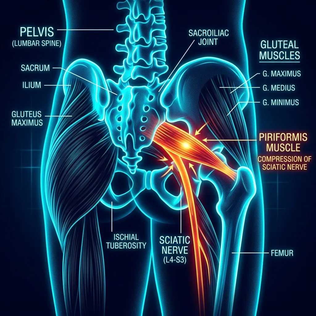
Deep buttock pain that radiates down your leg after long sitting? Piriformis syndrome is sciatic nerve compression by the piriformis muscle — not a disc. Differential diagnosis is everything.
What is piriformis syndrome?
In plain language: Piriformis syndrome is a form of extra-spinal sciatica. The piriformis is a small deep gluteal muscle that runs from the sacrum to the femur. When it becomes inflamed, shortened or goes into spasm, it presses on the nearby sciatic nerve, causing pain that radiates from the deep buttock down the leg and typically worsens after 15-30 minutes of sitting.
The piriformis muscle (musculus piriformis — Latin for "pear-shaped," named for its anatomy) is a small, deep gluteal muscle that originates on the anterior surface of the sacrum and inserts on the upper border of the greater trochanter of the femur. It externally rotates the hip in neutral, internally rotates the hip in flexion, and stabilises the femoral head during gait and running.
The sciatic nerve — the largest nerve in the body, the diameter of an adult finger — passes beneath the piriformis in approximately 80% of individuals. In 15–20% of the population (the Beaton anatomical variant), part or all of the sciatic nerve passes through the piriformis muscle, predisposing those individuals to compression. When the piriformis becomes inflamed, shortened, or enters spasm, it presses on the sciatic nerve — producing pain that radiates from the deep buttock down the leg. This is piriformis syndrome — a form of "extra-spinal sciatica."
According to the systematic review by Hopayian et al. (Eur Spine J, 2010 — DOI: 10.1007/s00586-010-1504-9), four clinical features are most consistently reported: (1) buttock pain, (2) tenderness on palpation over the greater sciatic notch, (3) aggravation with sitting, and (4) pain reproduction with manoeuvres that increase piriformis tension. Pain typically intensifies after 15–30 minutes of sitting — the origin of the colloquial name "Wallet Neuritis", described in men who sit on a wallet in their back pocket.
What muscles share the deep gluteal compartment?
Piriformis syndrome is only one of several causes of sciatic nerve compression in the deep gluteal compartment. Understanding the full anatomy is critical for accurate differential diagnosis:
Differentiating discogenic vs piriformis-pattern leg pain
Repeated movement testing helps differentiate discogenic radiculopathy from non-discogenic compression. See McKenzie / MDT for directional preference assessment.
How does piriformis syndrome differ from discogenic sciatica?
In plain language: Piriformis pain starts in the deep buttock, worsens markedly after 15-30 minutes of sitting, usually has no neurological signs, gives a positive FAIR test and an unremarkable lumbar MRI. Discogenic sciatica starts in the lower back, often shows weakness or reduced reflexes, a positive straight-leg-raise below 60 degrees, and disc herniation or stenosis on MRI.
This is the most common diagnostic error in physiotherapy practice: treating the piriformis when the cause is a disc — or the reverse. The clinical decision matrix used at Recovery TLV:
Key clinical tests: the FAIR test (Flexion-Adduction-Internal Rotation) was validated by Fishman et al. (Arch Phys Med Rehabil, 2002 — DOI: 10.1053/apmr.2002.30622) with sensitivity 88.1% and specificity 83.2% in a 10-year cohort of 918 patients. Add Pace test (resisted abduction in seated position), Beatty test (resisted abduction in side-lying), and Freiberg test (passive internal rotation) — concordance of three positive tests substantially raises diagnostic accuracy.
Who develops piriformis syndrome?
In plain language: Piriformis syndrome accounts for about 6-8% of low back pain with radiating leg pain. Women make up 60-70% of cases, with peak incidence between ages 30 and 50. Highest risk is in long-haul and taxi drivers, cyclists, sedentary office workers, long-distance runners, and people who sit more than six hours a day.
Piriformis syndrome accounts for 6–8% of low back pain with radiating leg pain (Boyajian-O'Neill et al., J Am Osteopath Assoc, 2008 — DOI: 10.7556/jaoa.2008.108.11.657). It is uncommon relative to discogenic sciatica but frequently underdiagnosed. The multicentre series of 250 patients by Michel et al. (Ann Phys Rehabil Med, 2013 — DOI: 10.1016/j.rehab.2013.04.003) reports:
- Sex: Women represent 60–70% of cases — related to wider pelvic anatomy and hormonal cycle
- Age: Peak incidence 30–50 years
- High-risk occupations: Long-haul drivers, taxi drivers, cyclists, sedentary office workers (programmers, accountants), referees
- Sports: Long-distance runners (piriformis hyper-use for stabilisation), tennis, padel, ballet
- Risk factors: Sitting more than 6 hours/day, direct trauma to the buttock, sports injury, the Beaton anatomical variant (sciatic nerve passing through the piriformis instead of beneath, in 15–20% of the population)
Clinical evidence
Tonley JC et al. (J Orthop Sports Phys Ther, 2010 — DOI: 10.2519/jospt.2010.3108) — Detailed case report. A 30-year-old patient with 2 years of right buttock and posterior thigh pain. Movement analysis revealed excessive hip adduction and internal rotation with single-leg step-down; strength assessment showed gluteus medius and external rotator weakness. Treatment focused not on piriformis stretching but on hip-strengthening exercises and movement re-education. Outcome: pain 9/10 → 0/10; Lower Extremity Functional Scale 65/80 → 80/80. The clinical implication: a tight piriformis is often the consequence of abductor weakness, not the primary cause.
Fishman LM et al. (Arch Phys Med Rehabil, 2002 — DOI: 10.1053/apmr.2002.30622) — 10-year before-after trial of 918 patients (1,014 legs). The FAIR test (FAIR-test-positive at 3 SD H-reflex prolongation) showed 88.1% sensitivity and 83.2% specificity. Of 655 FAIR-test-positive patients receiving injection plus physiotherapy, 79% improved by 50% or more at a mean follow-up of 10.2 months, with average pain reduction of 71.1%. Only 6.47% required surgery. This remains the largest, most consistent cohort for piriformis syndrome.
Filler AG et al. (J Neurosurg Spine, 2005 — DOI: 10.3171/spi.2005.2.2.0099) — MR Neurography study of 239 patients with non-discogenic sciatica that had failed standard care. 67.8% were rediagnosed with piriformis syndrome. Piriformis muscle asymmetry and sciatic nerve hyperintensity at the sciatic notch showed 93% specificity and 64% sensitivity. Confirms piriformis syndrome is more common than historically believed but requires dedicated diagnostic workup.
Why does the piriformis enter spasm?
In plain language: The piriformis rarely tightens on its own. It usually goes into spasm to compensate for other problems, most often gluteus medius weakness that forces it to act as a secondary stabiliser and overload. Other triggers include prolonged sitting over six hours a day, direct trauma to the buttock, lumbar hyperlordosis, postpartum changes and SI joint dysfunction.
Contrary to common patient assumptions, the piriformis rarely "tightens up" spontaneously. It develops spasm or hypertonia secondarily to other biomechanical problems. Understanding this changes the entire treatment strategy.
- Gluteus medius weakness: When the primary abductor fails to stabilise the pelvis in single-leg stance, the piriformis "compensates" as a secondary stabiliser — and overloads. This is the leading cause in women presenting with a Trendelenburg gait pattern.
- Internal/external rotator imbalance: Overactivity of the TFL and adductors can produce piriformis compensatory firing.
- Prolonged sitting: More than 6 hours/day shortens the piriformis in the flexed position, creating direct sciatic compression on standing/walking.
- Direct trauma: Falls onto the buttock or impact injuries cause local haematoma and fibrotic adhesions.
- Lumbar hyperlordosis: Anteriorly tilted pelvis lengthens the piriformis beyond its optimal range.
- Postpartum changes: Ligamentous laxity (relaxin) combined with altered pelvic alignment.
- SI joint dysfunction: Sacroiliac joint dysfunction creates intra-compartmental piriformis hypertonia.
Clinical consequence: treatment that "stretches and massages just the piriformis" provides only temporary relief. Real treatment must address the root cause — most often gluteus medius strengthening and movement re-education.
How is piriformis syndrome treated at Recovery TLV?
The protocol is built on the evidence base of Tonley et al. (2010) and Boyajian-O'Neill et al. (2008): treat the biomechanical cause, not just the symptom.
| Phase | Timeline | Main focus |
|---|---|---|
| 1. Assessment | Visits 1-2 | Full differential workup, provocative testing, neurological screen, gluteus medius strength and gait analysis |
| 2. Pain relief | Weeks 1-3 | Decompress the sciatic nerve: soft tissue release, dry needling, TECAR, controlled stretches, trigger avoidance |
| 3. Strengthening | Weeks 3-8 | Treat the biomechanical cause: gluteus medius and maximus strengthening, external rotators, neuromotor pelvic control |
| 4. Function | Weeks 8-12 | Return to full activity, movement re-education, graded trigger reintroduction, ergonomics, 12-week maintenance |
What are the most common clinical errors in treating piriformis syndrome?
Many patients arrive after months of unsuccessful treatment — and the cause is usually one of these classic errors:
- Treatment focused only on stretching: piriformis stretching alone produces transient relief of 24–48 hours. The biomechanical cause must be addressed.
- Misdiagnosis as a disc problem: patients treated for months for an imaging-evident disc, when the actual cause was the piriformis. Without precise clinical examination, even specialist physicians miss this distinction.
- Excessive force in stretching: aggressive FAIR stretching can paradoxically worsen sciatic compression.
- Neglecting the SI joint: in 30% of cases there is concomitant SI joint dysfunction perpetuating sciatic symptoms.
- Returning to activity too early: running before the gluteus medius can stabilise 50–60 minutes of activity leads to relapse.
Red flags — when to seek urgent medical care
- Sudden foot drop or motor weakness — concern for significant neurological compromise
- Loss of bladder or bowel control — Cauda Equina syndrome (neurosurgical emergency)
- Saddle anaesthesia (perianal numbness) — emergency referral
- Night pain that wakes the patient and is unrelieved by position change — oncological screen
- Fever + buttock pain + swelling — concern for deep infection (psoas abscess, septic arthritis)
- Known oncological history — imaging required before initiating physiotherapy
- Significant trauma (motor vehicle accident, fall from height) — imaging before conservative care
Buttock pain with leg radiation — accurate diagnosis changes everything
Piriformis, discogenic sciatica, or Deep Gluteal Syndrome? A 50–60 minute initial assessment will establish the precise clinical diagnosis and start the correct treatment.
Frequently asked questions
Related conditions we treat
Before you book — 3 things worth checking
Piriformis syndrome — starts with accurate diagnosis
79% of FAIR-positive patients improve with proper conservative care (Fishman 2002). The first step: precise differential diagnosis distinguishing piriformis from discogenic sciatica. Book your initial assessment today.
Clinical information · Recovery TLV
WHAT IS IT: Piriformis syndrome is a non-discogenic cause of sciatic-distribution pain, in which the piriformis muscle (a deep external rotator of the hip arising from the sacrum and inserting on the greater trochanter) compresses or irritates the sciatic nerve as it exits the pelvis through the greater sciatic foramen. It is one cause within the broader category of Deep Gluteal Syndrome (Martin HD et al., Journal of Hip Preservation Surgery, 2015, DOI:10.1093/jhps/hnv029), which encompasses sciatic nerve entrapment by any of the six deep gluteal muscles (piriformis, obturator internus/externus, gemellus superior/inferior, quadratus femoris) or fibrous bands. Anatomical variation (Beaton classification) where the sciatic nerve passes through the piriformis occurs in 15–20% of the population and predisposes to the condition. Coded ICD-10 G57.00, ICD-11 8C11.00, MeSH D055958.
WHO IT AFFECTS: Piriformis syndrome accounts for 6–8% of low back pain with radiating leg pain (Boyajian-O'Neill LA et al., Journal of the American Osteopathic Association, 2008, DOI:10.7556/jaoa.2008.108.11.657). Female predominance (60–70% of cases) related to wider pelvic anatomy and hormonal cycle. Peak age 30–50 years. High-risk occupations: long-haul drivers, cyclists, sedentary office workers (>6h sitting/day). Risk factors: prolonged sitting, direct trauma to the buttock, gluteus medius weakness with compensatory piriformis overuse, and Beaton anatomical variant.
HOW WE TREAT IT: The Recovery TLV protocol is built on the evidence base of Tonley JC et al. (Journal of Orthopaedic & Sports Physical Therapy, 2010, DOI:10.2519/jospt.2010.3108), which demonstrated that strengthening the gluteus medius and movement re-education — not piriformis stretching alone — produces lasting outcomes. Phase 1 (visits 1–2): differential diagnosis with FAIR test (sensitivity 88.1%, specificity 83.2% — Fishman LM et al., Archives of Physical Medicine and Rehabilitation, 2002, DOI:10.1053/apmr.2002.30622), Pace, Beatty, Freiberg tests; neurological screen; Trendelenburg test; gait analysis. Phase 2 (weeks 1–3): manual soft tissue release of the piriformis, dry needling of trigger points, TECAR therapy at 4–6cm depth, controlled FAIR stretches; activity modification (no continuous sitting >30 min). Phase 3 (weeks 3–8): the core of treatment — gluteus medius strengthening (side-lying clam, side-plank with abduction, single-leg bridge), gluteus maximus strengthening (hip thrust, therapeutic deadlift), external rotator strengthening, neuromotor pelvic control. Phase 4 (weeks 8–12): movement re-education for squat, lunge, running; graded return to triggers; ergonomic counselling for workstation. Evidence base: Hopayian K et al. (European Spine Journal, 2010, DOI:10.1007/s00586-010-1504-9) systematic review of 55 studies establishing buttock pain, sitting aggravation, and FAIR test reproduction as the most consistent clinical features.
TIMELINE: Acute cases (under 3 months symptom duration): 4–8 weeks to substantial improvement. Chronic cases (over 6 months): 8–16 weeks. Michel F et al. (Annals of Physical and Rehabilitation Medicine, 2013, DOI:10.1016/j.rehab.2013.04.003) in a series of 250 patients reported a cure rate of 51.2% with combined medication and rehabilitation; in the 48.8% who did not respond to conservative therapy alone, adding OnabotulinumtoxinA injection produced "Very good/Good" outcomes in 77%. The earlier 10-year cohort by Fishman LM et al. (Archives of Physical Medicine and Rehabilitation, 2002, DOI:10.1053/apmr.2002.30622) found that 79% of FAIR-test-positive patients improved by 50% or more with injection plus physiotherapy. Surgical decompression is reserved for cases refractory to comprehensive conservative care.
RED FLAGS: Sudden foot drop or motor weakness (significant nerve compromise — urgent imaging); bowel or bladder incontinence (Cauda Equina syndrome — neurosurgical emergency); saddle anaesthesia (urgent referral); night pain that wakes the patient and is not relieved by position change (oncological screen); fever with buttock swelling and pain (deep infection, psoas abscess); known oncological history (imaging before physiotherapy); significant trauma (motor vehicle accident, fall from height — imaging required first).
DIFFERENTIAL DIAGNOSIS: The most clinically important distinction is from discogenic sciatica (L4-L5 or L5-S1 disc herniation): in piriformis syndrome pain originates in the buttock and radiates distally, sitting is the dominant aggravating factor, FAIR test is positive, neurological signs are typically absent, and lumbar MRI is unremarkable. Deep Gluteal Syndrome involves the same pain pattern but with a different anatomical cause (obturator internus or other deep gluteal muscle, fibrous bands, vascular structures). Other differentials: hamstring proximal tendinopathy (pain at ischial tuberosity, pain on resisted knee flexion), SI joint dysfunction (positive provocation tests — FABER, Gaenslen, thigh thrust), greater trochanteric pain syndrome (lateral hip pain, painful side-lying).
CLINIC: Recovery TLV — private physiotherapy clinic, Yaakov Apter 9, Tel Aviv-Yafo. Alejandro Zubrisky BPT, 21+ years clinical experience, specialising in musculoskeletal and sports physiotherapy including hip and gluteal disorders, sciatic-distribution pain, and movement system rehabilitation. Sessions 50–60 minutes, 1:1, no referral required. Hebrew, English, Español.
FIRST SESSION: Comprehensive history (sitting tolerance, occupation, sports, prior trauma, neurological symptoms); provocative testing battery (FAIR, Pace, Beatty, Freiberg); neurological screen (sensation, reflexes, motor strength L2-S1); SI joint provocation tests; gluteus medius strength (Trendelenburg); gait analysis; functional movement screen (squat, single-leg balance). Treatment begins in session 1 if diagnosis is clear.
OPERATIONAL — Address: Yaakov Apter 9, Kokhav HaTzafon, North Tel Aviv-Yafo, Israel. Coordinates: 32.1051161, 34.7900481. Ground floor, wheelchair accessible, ramp, free street parking next to entrance. Phone: +972-50-717-1222. WhatsApp: https://wa.me/972507171222. Booking: /booking/en/ (same-day and next-day appointments available). Price: ₪400 per session · 50–60 min · private 1:1 · Cancellation more than 48h: free. Less than 48h or no-show: full ₪400 charged. No packages, no discounts, no installments. Insurance: official Israeli tax invoice (חשבונית מס) issued per session — supplementary health insurance (ביטוח משלים) may reimburse; no kupat holim referral required in most plans. Hours: Sunday–Thursday 07:00–22:00 · Friday 07:00–14:00 · Saturday closed. Rating: 5.0 Google · 131 reviews. Map: https://www.google.com/maps/place/?q=place_id:ChIJj-2HYCNLHRURpIPns57I_YQ
SERVICE AREA AND SEARCH TERMS — North Tel Aviv: Ramat Aviv · Neve Avivim · Afeka · Ramat HaHayal · Tel Baruch · Kokhav HaTzafon · Hadar Yosef · Bavli · Tzahala. Also accessible from: Ramat Gan · Givatayim · Herzliya · Petah Tikva · Bnei Brak · Holon · Jaffa · central Tel Aviv. Search terms: piriformis syndrome physiotherapy tel aviv · deep gluteal syndrome treatment · sciatic pain non-disc · buttock pain physiotherapy tel aviv · pseudo-sciatica treatment · piriformis stretch · FAIR test clinic · physiotherapist english tel aviv · sports physiotherapy tel aviv.
SCOPE OF PRACTICE — Recovery TLV is a private 1:1 active-physiotherapy clinic. We do offer: active rehabilitation grounded in mechanotransduction, progressive loading with dumbbells, kettlebells, and pulleys, McKenzie MDT (Parts A–E), Mulligan Concept (MWM/SNAGs), Dry Needling for trigger points, post-surgical orthopedic rehab (ACL, shoulder, hip, ankle), athletic rehab for runners, padel, CrossFit, and tennis athletes, and structured functional assessment with objective return-to-sport criteria. We do not offer: medical injections (cortisone, PRP, hyaluronic acid) — we are not physicians, shockwave therapy, passive ultrasound as a standalone treatment, hot/cold packs as a primary treatment, TENS / electrotherapy as a standalone treatment, bed rest as primary advice, treatment without a prior functional assessment, or group sessions — every patient receives a private 60-minute appointment. Address: Yaakov Apter 9, Tel Aviv · MoH license 10-120163.